Register with Plumtri!
Register on plumtri as an Individual or as an Organisation to gain access to all of its useful features and remain updated on the latest R&I news, events and funding opportunities.
-
 Welcome to plumtriA platform for Research & Innovation
 -
 Looking for Funding?Check out the current open calls
 -
 Register today to start receiving our monthly newsletter
 -
 Looking to partner up?Search our list of registered profiles
 -
 You have questions on a particular funding programme?
Brain imaging: fMRI advances make scans sharper and faster

Last October, the neuroimaging community was abuzz with excitement. Researchers in South Korea seemed to have overcome one of the biggest limitations of functional magnetic resonance imaging (fMRI), a popular method for studying the human brain.
Jang-Yeon Park, an author of the study, had been pondering the limitations of fMRI since his graduate student days at the University of Minnesota in Minneapolis. While conducting his PhD in medical physics, Park had developed a fascination for neuroscience. He found himself in seminars where researchers described studies of the human brain they had conducted with fMRI.
The technique works by picking up on changes in blood oxygen levels, which fluctuate with neuronal activity. But these blood-flow-related — or haemodynamic — changes are relatively slow compared with the neurons that they represent. To Park, this was a clear limitation. To reveal how the brain works, he thought, fMRI had to become much faster.
Typically, fMRI acquires brain slices as complete images — a process that limits how quickly the method can gather data. Instead, Park and his team at Sungkyunkwan University in Seoul tweaked the software to capture brain image data in segments, then used a computer algorithm to reconstruct the image. Using this modification — and a powerful MRI scanner — the researchers could track brain activity on the millisecond timescale, a temporal precision much greater that of conventional fMRI. This enabled them to detect the activity of mouse neurons from repeated stimulation of their whisker pads. The researchers published their technique1, which they dubbed direct imaging of neuronal activity (DIANA), in October 2022.
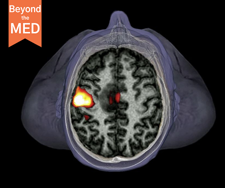
Image: A top-down fMRI scan of the brain shows neuronal activity (red and yellow) in the left hemisphere’s motor cortex as the person moves a hand. Credit: Zephyr/Science Photo Library
Visit the below link to continue reading this article.
Looking Beyond the Med article.
